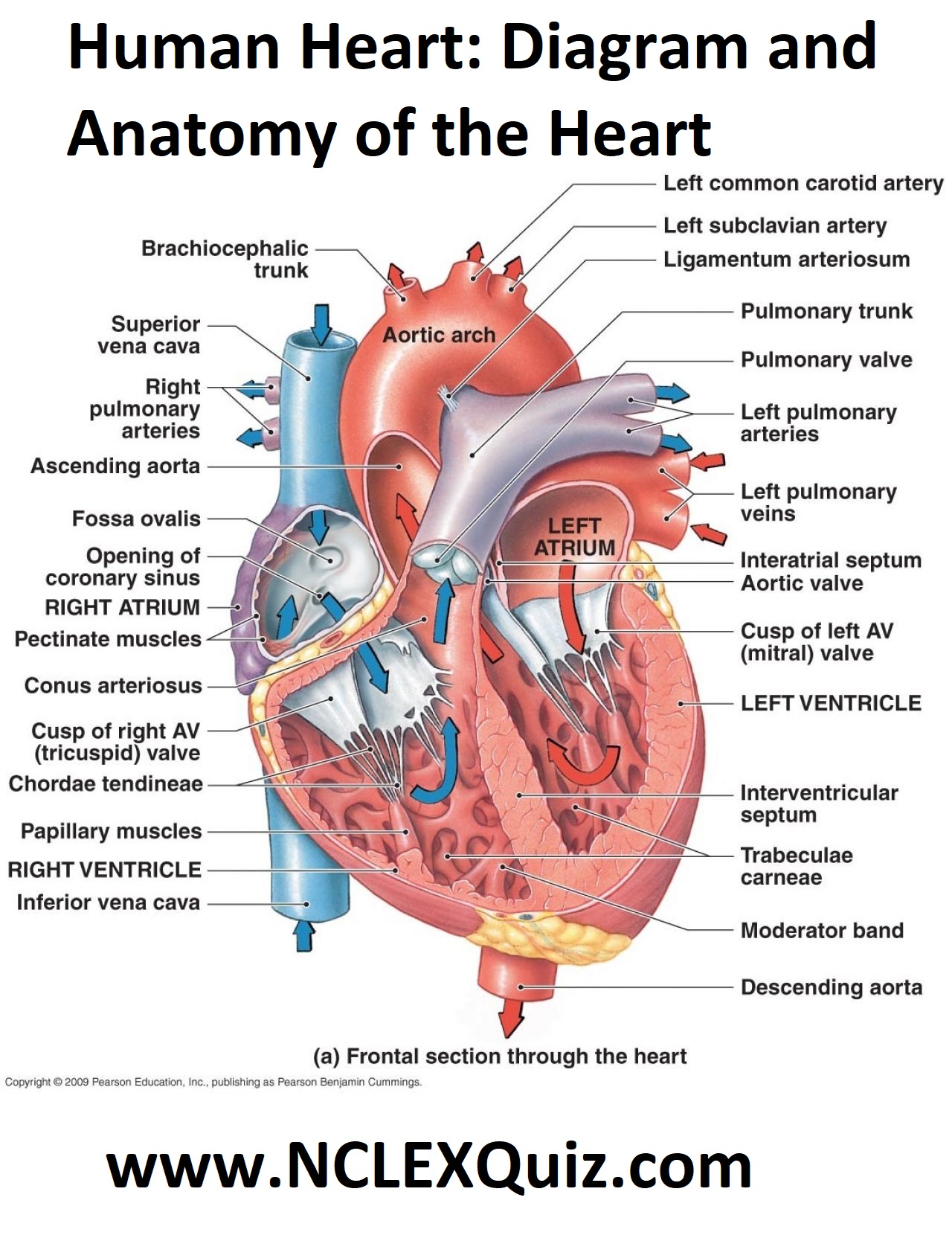
Human Heart: Diagram and Anatomy of the Heart
Internal Anatomy of the Heart
The human heart is a muscular organ that pumps blood throughout the body. It is located in the center of the chest and is about the size of a fist. The heart is divided into four chambers: two atria and two ventricles. The atria receive blood from the body and the veins, and the ventricles pump blood out to the body and the arteries.
- Right atrium: The right atrium receives blood from the body through the superior and inferior vena cavae.
- Left atrium: The left atrium receives blood from the lungs through the pulmonary veins.
- Right ventricle: The right ventricle pumps blood to the lungs through the pulmonary trunk.
- Left ventricle: The left ventricle pumps blood to the rest of the body through the aorta.
The chambers of the heart are separated by two septa: the interatrial septum and the interventricular septum. These septa prevent blood from mixing between the different chambers of the heart.
The heart also has four valves: the tricuspid valve, the mitral valve, the aortic valve, and the pulmonary valve. These valves open and close to control the flow of blood through the heart.
Heart Diagram: Right/left Atria, Right/left Ventricles, Pulmonary Trunk, Aorta, Superior/inferior Vena Cavae, Pulmonary Veins, Coronary Sinus, Right/left Atrioventricular valves (tricuspid + bicuspid), Chordae Tendinae, Interatrial Septum, Interventricular Septum, Aortic and Pulmonary Semilunar Valves, Coronary Arteries and Cardiac Veins.

Leave a Reply