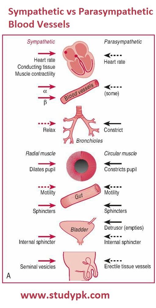
Comparison of Sympathetic and Parasympathetic Effects on Blood Vessels
Nervous System Physiology Sympathetic and Parasympathetic Nervous System
In nursing education guidelines, it is important to understand the difference between sympathetic and parasympathetic blood vessels. Under sympathetic stimulation, blood vessels constrict, leading to an increase in pressure. In contrast, the parasympathetic division directs the body towards a “rest and digest” mode, which generally decreases heart rate and blood pressure.
It is important to note that major arteries and precapillary arterioles are innervated by sympathetic nerves, while other vessels, such as venules, capillaries, and collecting veins, are rarely innervated. Blood vessels can receive innervation from three main classes of neurons: sympathetic vasoconstrictor neurons, sympathetic or parasympathetic vasodilator neurons, and sensory neurons that mediate vasodilation.
Sympathetic stimulation increases metabolic activity, leading to vasodilation. This results in the relaxation of the muscles, leading to the widening of blood vessels and affecting the luminal blood flow. The primary effect of sympathetic stimulation in the arterioles is to produce arteriolar constriction, especially in the kidneys, spleen, and skin. However, there are important exceptions such as the heart, bronchial circulation, and the brain, where sympathetic stimulation produces arteriolar dilation.

Leave a Reply