Magnetic resonance imaging (MRI) is an important tool for diagnosing and evaluating diseases. The process can be summarized as induction of protons by a magnetic field, followed by excitation with radio frequency pulses and subsequent readout with receiver coils.
One report from the Joint Commission showed burns are the most common injuries in the MRI suite. The most common objects to undergo significant heating are wires and leads, pulse oximeter sensors and cables, and safety pins and metal clamps. Others include drug-delivery patches (which may contain a metallic coil), cardiorespiratory cables, and tattoos (which may contain iron oxide pigment).
In another study, MRI-related incidence was significantly higher in inpatient settings compared with outpatient centers (74% vs 29%).Additionally, medication safety was reported as the third-most frequent safety incident.
Transdermal patches are self-contained discrete dosage forms that deliver drugs through the skin to the systemic circulation.Their structural elements include:
Backing layer,
Drug layer or reservoir,
Adhesive layer, and
Protective or release liner.
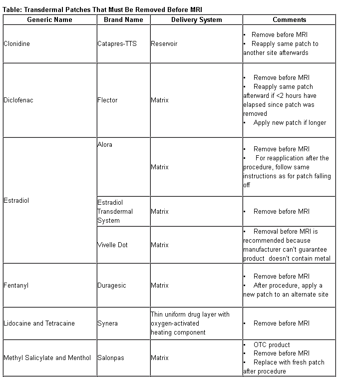
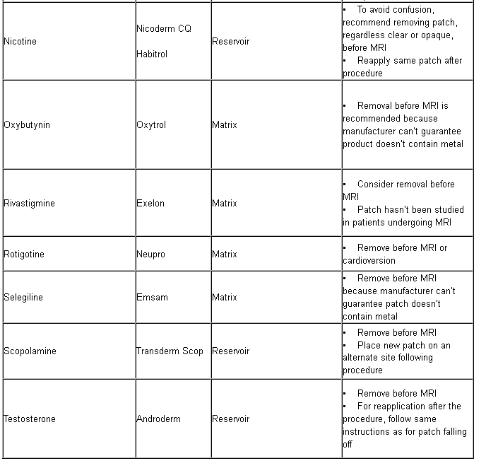
Some transdermal patches containing aluminum or other metal in their nonadhesive backing shouldn’t be worn during MRI because of skin burn risk. Health care professionals should advise patients wearing medication patches about procedures for proper removal and disposal before MRI and replacement afterwards.The FDA is currently reviewing transdermal products containing metal (Table) to ensure they carry a warning about burn risk during MRI.


Tobias Gilk says
This article appears to have been copied, almost in its entirety, from Pharmacy Times (http://www.pharmacytimes.com/contributor/alexander-kantorovich-pharmd-bcps/2016/08/transdermal-patches-that-must-be-removed-before-mri) without any attribution. This appears to be intellectual property misappropriation.